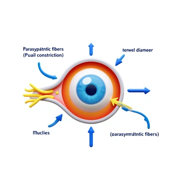
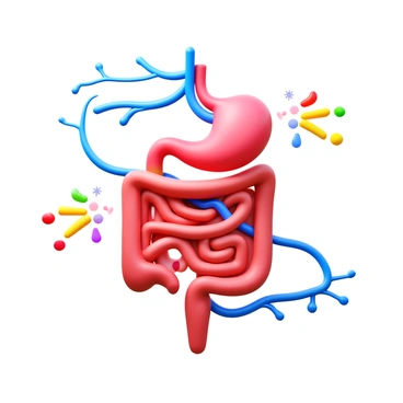
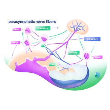
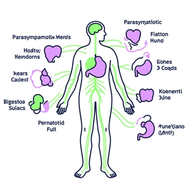
Label structures parasympathetic division

An illustration depicting the branching of nerves in the parasympathetic division of the autonomic nervous system. The image includes a detailed network of nerve fibers with a soft color palette, illustrating the pathways of the parasympathetic nerves. Each branch is clearly defined, showing the connections to various organs and tissues. Labels on the nerves indicate specific anatomical locations, enhancing clarity and educational value. The overall composition emphasizes the intricate system of the parasympathetic nervous system and its role in bodily functions.A 3D icon depicting neurotransmitters in a neural network. The neurotransmitters are represented as small, vibrant spheres in various colors, such as blue, green, and yellow. These spheres are shown transmitting signals between neurons in the parasympathetic nervous system. The neurons are illustrated with elongated shapes and branching dendrites. The background features a subtle gradient that enhances the focus on the neurotransmitter activity.A detailed illustration of the brainstem, highlighting its role in the parasympathetic division of the autonomic nervous system. The brainstem is depicted with clear anatomical features, showcasing the medulla oblongata, pons, and midbrain. Include labeled arrows indicating the pathways through which the vagus nerve branches extend from the brainstem to various organs. Surround the brainstem with icons representing its functions, such as a heart for heart rate regulation, a stomach for digestive processes, and lungs for respiratory control. The overall composition emphasizes the connection between the brainstem and the parasympathetic nervous system, illustrating its importance in maintaining homeostasis.A modern pastel illustration showing a human silhouette in a relaxed seated position surrounded by soft, flowing lines representing calmness. The silhouette is filled with gentle shades of blue and green, symbolizing tranquility. Scattered around the figure are visual elements such as a heart, a brain, and soothing waves, each portrayed in light pastel colors. Light rays emanate from the silhouette, creating an aura of peace. A small icon of a leaf is present to signify renewal and relaxation.A 3D icon representing the balance between the "sympathetic" and "parasympathetic" nervous systems. On one side, the "sympathetic" system is illustrated with vibrant red tones and dynamic, angular shapes, symbolizing alertness and activity. On the opposite side, the "parasympathetic" system is depicted in calming blue hues and smooth, flowing forms, representing relaxation and rest. In the center, a scale balances these two systems, emphasizing their interaction and equilibrium.An illustration depicting the parasympathetic nervous system's innervation of the heart. In the center, there is a detailed anatomical heart, showing major arteries and veins. Surrounding the heart are nerve fibers represented in soft pastel colors, highlighting the vagus nerve extending from the brainstem down towards the heart. A labeled diagram indicates the pathways of the nerve fibers, with arrows showing the direction of impulse transmission. Alongside, there are illustrations of neurotransmitters in different pastel shades, demonstrating their role in slowing the heart rate. The background is subtly colored to enhance the clarity of the heart and nerve structures.A serene scene illustrates the concept of "rest and digest." A peaceful indoor environment features a comfortable armchair where a person is seated, surrounded by soft cushions. On a nearby table, a bowl of fresh fruits, including apples and bananas, sits invitingly. A gentle light glows from a lamp, and a stack of books is neatly arranged beside the chair. A small potted plant with lush green leaves adds a touch of nature to the space. The overall atmosphere suggests relaxation and calmness, embodying the essence of tranquility and digestive wellness.A detailed illustration depicting the "medulla oblongata" at the center of the image, showcasing its prominent position within the human brain. The medulla is colored in light gray, with highlighted areas in soft blue to indicate its involvement in parasympathetic regulation. Surrounding the medulla are simplified, labeled lines connecting it to various body systems, such as the heart, lungs, and digestive organs. Each line leads to pastel-colored icons representing the functions controlled by the parasympathetic nervous system, including a heart symbol, a pair of lungs, and a stomach. The background is a gentle gradient of pastel colors, enhancing the clarity of the central elements.A detailed illustration of the human brain focusing on the hippocampus. The hippocampus is depicted in a prominent position, with clear labeling. Thin lines connect the hippocampus to areas of the brain associated with parasympathetic activities, such as the vagus nerve and brainstem regions. The vagus nerve is illustrated as a long, winding path extending from the brainstem to various organs, emphasizing its role in the parasympathetic nervous system. Additionally, arrows indicate the flow of signals between these areas, highlighting their interactions and functions. The overall composition is organized to clearly show the anatomical connections and pathways.A diagram illustrating the "parasympathetic effects on the respiratory passages" is displayed. The central focus is on the respiratory system, featuring bronchi and bronchioles. Arrows indicate the pathways of nerve signals originating from the parasympathetic nervous system. The airways are depicted in a slightly constricted state, demonstrating bronchoconstriction. Surrounding the diagram are labeled sections with key terms such as "bronchoconstriction" and "increased mucus secretion" highlighted in contrasting colors. The overall diagram is arranged in a clear, logical manner, facilitating easy understanding of the physiological processes.An educational illustration displaying the "vagus nerve" in a detailed manner. The image shows the path of the nerve, originating from the "brain" and tracing down through the neck and torso, with clear labels along the route. The nerve is represented as a distinct, wavy line, transitioning in color from light blue near the brain to deeper blue as it travels downwards. Surrounding the nerve are simplified illustrations of key organs it interacts with, including the "heart," "lungs," and "stomach," each clearly labeled to indicate their relation to the vagus nerve. The background is a soft, pastel color that enhances the visibility of the nerve and organs.A diagram illustrating the "parasympathetic connections to thoracic organs." The central focus is a stylized representation of the human torso, with highlighted thoracic organs such as the heart and lungs. Various pathways are depicted in soft pastel colors, connecting the brainstem and the vagus nerve to these organs. Arrows indicate the direction of nerve signals, and labeled lines show the specific connections to the heart, lungs, and other thoracic areas. Each organ is visually distinct and clearly marked, while the background maintains a simple, light pastel tone to emphasize the diagram's elements.An illustrated map of the brain showcases various regions, with a clear focus on the areas highlighting the origin of the parasympathetic nerves. The map includes detailed labels for each brain region, using different pastel colors for contrasting sections, such as light blue for the brainstem and soft green for the cranial nerves. Small icons, like nerve endings and neurons, are incorporated around the highlighted areas to represent the nervous system visually. Alongside the map, there are arrows pointing to the origin points of the parasympathetic nerves, all presented within a clean and organized layout.A close-up illustration of a synapse in the parasympathetic division. The synaptic cleft is visible between two neurons. One neuron has branched dendrites extending towards the synapse, while the other neuron features a rounded synaptic terminal with vesicles. Neurotransmitters are depicted as small circles being released into the synaptic cleft. The background includes additional neural structures such as axons and myelin sheaths, adding context to the synapse's location within the nervous system.An illustration depicting the mechanism of parasympathetic control of pupil constriction. The image shows a close-up view of an eye with the iris visibly constricting, demonstrating the reaction. Arrows indicate the nervous pathways involved in this process, highlighting the connection between the brain and the eye. The pupil is shown in a smaller diameter, emphasizing the constriction. Labels in the image explain the different components involved, such as the parasympathetic fibers and the iris muscles.A diagram illustrating the autonomic nervous system, prominently featuring the parasympathetic division. The diagram includes labeled pathways connecting the brain to various organs such as the heart, lungs, and digestive system. Key components, such as the vagus nerve, are highlighted. The parasympathetic division is marked in a distinct color to emphasize its role in relaxation and bodily functions. Clear arrows indicate the direction of signals from the brain to the organs. The overall layout is organized and easy to understand, providing a clear visual representation of the system.A 3D icon representing the "parasympathetic influence on intestinal motility". The icon features a detailed depiction of the digestive system, highlighting the intestines with arrows symbolizing movement. The parasympathetic nervous system is represented by a network of nerves in blue color, connecting to the intestines. The intestines are shown in a relaxed state, with subtle movement indicated by soft waves. Additionally, there are graphical representations of neurotransmitters depicted in vibrant colors, illustrating their role in enhancing motility.A close-up view of neuron pathways in the parasympathetic division. The image features a network of neurons with visible cell bodies and extending axons. Each pathway is intricately detailed, showcasing synapses and dendrites connecting in a complex arrangement. Labels are positioned clearly along the pathways, identifying key components such as "axon," "dendrite," and "synapse." The overall composition highlights the structure and connections of the parasympathetic nervous system, emphasizing the flow of information within this neural network.A detailed illustration of the pathway of the pelvic splanchnic nerves within the parasympathetic division. The image shows the spinal cord with the pelvic splanchnic nerves emerging from the sacral region. These nerves travel downward and branch out toward various pelvic organs. The pathway includes the pathways leading to the bladder, rectum, and reproductive organs. Arrows indicate the direction of the nerve pathways, and labels mark the spinal roots, ganglia, and target organs. The overall structure is clear and informative, highlighting the connections and routes taken by the pelvic splanchnic nerves.A detailed illustration of a network diagram showcasing "parasympathetic nerve fibers." The diagram features intricate lines connecting various nodes that represent different organs and areas of the body influenced by these nerve fibers. Emphasis is placed on the connectivity between the brainstem and the heart, lungs, and digestive organs, using different colors to highlight these pathways. Labels are placed prominently next to each node, with terms like "heart," "lungs," and "digestive organs" clearly visible. The overall layout is clean and organized, allowing for easy comprehension of the parasympathetic nervous system's structure.An illustration depicts the parasympathetic nerves connecting to various organs in the human body. In the center, a detailed anatomical figure shows the silhouette of a human body. The parasympathetic nerves are represented in a green-and-purple outline, extending from the brain and spinal cord to different organs. The connections reach the heart, which is shown beating, the lungs represented in a full shape, and the digestive organs, including the stomach and intestines, illustrated with lines indicating nerve pathways. The kidneys and bladder also have visible connections, all demonstrating the network of the parasympathetic nervous system clearly.A modern pastel illustration depicting a human head with highlighted facial nerves. The facial nerves are shown in bright colors, with arrows indicating their pathways. Surrounding the head are icons representing various parasympathetic actions, such as a water droplet symbolizing tear production, a heart symbol indicating slowed heart rate, and a digestive system graphic representing increased salivation and digestion. Each icon is connected to the respective facial nerve pathways to illustrate the connections and functions.A detailed illustration of parasympathetic ganglia. The image features multiple ganglia, including their anatomical locations, labeled clearly. Each ganglion is colored distinctly for easy identification. The surrounding neurons are depicted, connecting to various organs. Informative labels indicate the names of the ganglia and their functions. The image is presented in a clear, educational format.A simple diagram illustrating parasympathetic stimulation of the salivary glands. In the center, there is a large salivary gland labeled clearly. Arrows originating from a brain region, labeled "Brainstem," point towards the salivary gland, indicating the signal of stimulation. Surrounding the salivary gland are various smaller structures, such as nerve fibers, represented as thin lines leading to the gland. Additionally, small droplets are drawn near the gland to symbolize saliva production. The entire composition is clean and organized for clarity.A diagram illustrating the flow of nerve impulses in the parasympathetic division of the autonomic nervous system. The diagram should include "preganglionic neurons" originating from the brain and sacral spinal cord, extending towards "ganglia" located near or within target organs. The diagram should also show "postganglionic neurons" that connect to various organs, including the heart, lungs, and digestive system. Arrows should indicate the direction of impulse transmission, highlighting the pathway of nerve signals as they travel through the parasympathetic system. Color coding can be used to differentiate between preganglionic and postganglionic pathways.A split illustration compares the sympathetic and parasympathetic divisions of the autonomic nervous system. On the left side, the sympathetic division is depicted with a silhouette of a human body showing highlighted areas such as the adrenal glands. There are arrows indicating increased heart rate and energy levels. Sweat glands and dilated pupils are also illustrated to represent the body's response to stress. 

On the right side, the parasympathetic division is shown with a similar human silhouette but with highlighted areas such as the heart and digestive organs. Arrows indicate decreased heart rate and enhanced digestion. The illustration includes details like relaxed facial muscles and constricted pupils, representing the body's state of rest. The two sides contrast the features and functions of each division clearly.A modern pastel illustration featuring a detailed chart illustrating the cranial nerves involved in the parasympathetic system. The chart includes labeled diagrams of cranial nerves III (Oculomotor), VII (Facial), IX (Glossopharyngeal), and X (Vagus), each connected by colorful arrows showing their pathways. Surrounding the chart are icons representing various functions of these nerves, such as a pair of eyes for nerve III, a smiling face for nerve VII, a throat for nerve IX, and a heart for nerve X. The overall layout is clean and organized, with a soothing color palette that enhances readability.
В сообществе ничего не найдено