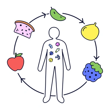
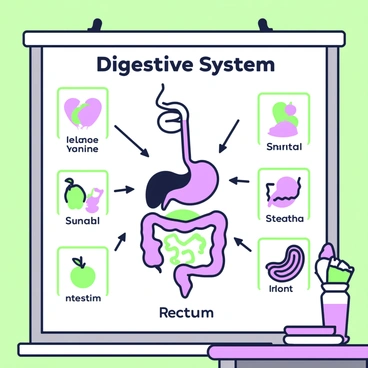
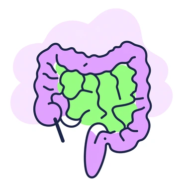
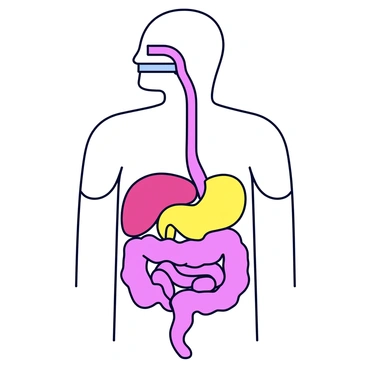
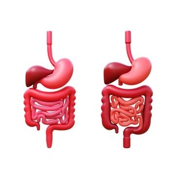
Human digestive system

A variety of colorful fruits and vegetables arranged artfully on a wooden table. Bright green kale leaves are placed alongside vibrant red tomatoes, orange carrots, and yellow bell peppers. Sliced fruits such as purple figs, green kiwi, and juicy watermelon are also included, showing their rich textures and colors. In the background, there is a softly blurred glass of water with lemon slices floating on top. The entire composition evokes a sense of freshness and vitality, emphasizing the theme of digestive health. The fruits and vegetables are depicted with detailed textures, highlighting their natural brilliance.A group of enzymes depicted as small, cartoon-style workers. Each enzyme has a unique appearance, with different colors representing various types. They are actively engaging with colorful food particles, such as a yellow piece of cheese, green broccoli, and red tomatoes. The enzymes are using tools like tiny hammers and shovels to break down the food particles into smaller pieces. In the background, there are simple, stylized shapes representing a kitchen setting.A representation of various food items, including a slice of bread, a piece of fruit, and a vegetable, arranged in a circular layout. Arrows point from each food item towards a simplified outline of a human body in the center. Inside the outline, small colorful icons represent different nutrients like vitamins, proteins, and carbohydrates being absorbed into the body. The background is a solid color to emphasize the main elements.An anatomical chart displaying the detailed parts of the "digestive system." The chart includes labeled structures such as the "esophagus," "stomach," "small intestine," "large intestine," "liver," "pancreas," and "gallbladder." Each organ is illustrated with accurate anatomical features in a clear, organized layout. The background of the chart is neutral to enhance visibility and comprehension of the digestive system's components.An educational poster is displayed on a classroom wall, illustrating the digestive system. At the center of the poster, there is a detailed diagram of the human digestive tract, featuring the mouth, esophagus, stomach, small intestine, large intestine, and rectum. Each organ is labeled clearly with text in quotes. Surrounding the central diagram are smaller images of various foods like fruits, vegetables, and grains, showing how they interact with the digestive organs. Arrows in green and purple trace the path of food through the system, leading from the mouth to the rectum. Additional text boxes in quotes provide information about the functions of each part of the digestive system.An illustration depicting natural foods being digested at different stages. At the top left, there is a whole carrot, vibrant and intact. To the right of the carrot, a half-sliced apple reveals its core and seeds. Below these, a piece of bread is shown partially broken apart, with soft interior visible. Progressing to the right, there is a bowl with a mixture of mashed fruits, representing a further stage of digestion. Finally, at the bottom right, there are simple nutrient shapes, such as circles and squares, that symbolize fully digested nutrients ready for absorption. Each stage is clearly labeled with arrows connecting them in a sequential flow.A close-up view of a section of the small intestine. The walls are detailed, showcasing the intricate folds and texture. Villi are prominently displayed, extending from the surface in various sizes. Each villus is slender and elongated, maximizing surface area for nutrient absorption. The background is a soft gradient, ensuring the focus remains on the villi and the structure of the intestine.A detailed anatomical illustration of the "liver" featuring its distinct lobes and textures. The "gallbladder" is prominently displayed beneath the liver, showing its curved shape. "Bile ducts" are clearly outlined, connecting the liver and gallbladder, with bile flowing through them. The entire structure is presented in a 3D perspective, emphasizing depth and detail.A detailed 3D render of a human digestive system with a strong focus on the intestines. The intestines are vividly colored, showcasing the small intestine coiling intricately and the large intestine appearing thicker and more robust. Surrounding the intestines, there are various labeled anatomical features including the stomach, liver, and pancreas, each depicted in a realistic manner. The background is a subtle gradient to enhance the focus on the intestines and surrounding organs.A human stomach actively digesting food. The stomach walls are slightly stretched to show the contents inside. Visible gastric juices are present, depicted as a translucent greenish fluid mixing with partially digested food. The interior has textures representing the digestion process, with small bubbles indicating the chemical reactions taking place.A detailed interior view of human intestines, highlighting the intricate structure of the intestinal walls, with folds and villi visible. Various types of bacteria, represented in different colors and shapes, are interacting with the intestinal lining. Some bacteria are clustered in groups, while others are swimming freely in the intestinal fluid. The scene captures a vibrant and dynamic ecosystem, showcasing the diversity of the gut microbiome and its close relationship with the intestines.A detailed chart with various "digestive enzymes" listed on the left side. Each enzyme has a corresponding description of its "function" on the right side. The chart features clear rows and columns, with well-defined borders. There are icons representing each enzyme, such as a small depiction of a "protease" enzyme for protein digestion and an image of "amylase" for starch breakdown. The background is plain to enhance readability, and the entire layout is organized and easy to follow.A cross-sectional view of the human abdomen showcasing the digestive organs. The detailed illustration features the stomach positioned in the center, with the small intestine coiled around it. The large intestine is visible, curling around the lower part of the abdomen. The liver is prominently displayed on the right side, while the pancreas is depicted beneath the stomach. Surrounding these organs are veins and arteries, with a subtle shading to indicate different structures. The entire view is presented with clear labels for each organ, enhancing the educational aspect of the illustration.A detailed view of the small intestine, showcasing the process of nutrient absorption. The interior of the small intestine is prominently displayed, with numerous villi, tiny finger-like projections that line the intestinal walls. Each villus is depicted with a rich surface texture, illustrating how nutrients are absorbed into the bloodstream. Various nutrients, such as glucose, amino acids, and fatty acids, are visually represented flowing into the capillaries within the villi. The surrounding environment includes digestive enzymes and partially digested food particles, highlighting the digestive process. Clear labels point to key components such as "villi," "capillaries," and "nutrients," enhancing the educational aspect of the image.A close-up view of the human stomach showcasing its intricate structure. The image features the muscular layers of the stomach walls in various shades of pink and beige, clearly delineating the outer serosa, the muscularis layers, and the inner mucosa, which displays a textured surface. The inner lining is highlighted with folds known as rugae, exhibiting a subtle sheen. Surrounding the stomach are labeled sections indicating the esophagus and small intestine, providing context for the stomach's location in the digestive system.A human body depicted in a transparent style, revealing the internal organs. The digestive system is prominently featured, showcasing the esophagus, stomach, small intestine, and large intestine in a detailed manner. Each organ is represented with clear outlines and vibrant colors, allowing for easy identification. The organs are connected by ducts and tubes, illustrating the flow of food. Small details, such as the texture of the organs and their natural shapes, enhance the visual representation.An illustration showing a detailed view of the pancreas positioned next to the small intestine. The pancreas is depicted with ducts leading to the small intestine, where small droplets representing enzymes are being released. The small intestine is shown with a textured surface, illustrating its coiled structure. Additional anatomical labels highlight the pancreas and the small intestine for educational purposes.A flowchart illustrating the pathway of food through the digestive system. The flowchart starts with the word "Mouth" at the top, leading to "Esophagus" as the next step. Arrows connect each stage, moving down to "Stomach," followed by "Small Intestine," "Large Intestine," and ending with "Anus" at the bottom. Each section of the flowchart contains labels that explain the process, such as "Chewing," "Swallowing," "Digestion," "Absorption," and "Excretion." The entire flowchart is surrounded by decorative elements that signify the digestive process.A detailed side-by-side comparison of a human digestive system and a cat's digestive system. On the left, the human digestive system is depicted with clearly labeled organs including the esophagus, stomach, small intestine, large intestine, and rectum. On the right, the cat's digestive system features similar organs, proportionally adjusted for size, with labels for the esophagus, stomach, small intestine, cecum, large intestine, and rectum. Both systems are colored in realistic shades, showcasing the differences in length and structure, such as the cat's shorter small intestine and presence of a cecum. Each organ is rendered with a 3D effect to enhance depth and dimension.A modern pastel illustration depicting a detailed human digestive system. The organs, including the stomach, intestines, and liver, are rendered in soft, pastel colors, emphasizing their shapes and functions. Glowing light radiates from the digestive organs, creating a sense of vitality and health. Surrounding the digestive system, there are fresh fruits and vegetables, such as apples, carrots, and leafy greens, enhancing the theme of nourishment. The background is a gentle blend of pastel hues, which complements the illustration and emphasizes the focus on the digestive system.A detailed diagram showcasing the "large intestine" in a vertical format. The large intestine is illustrated in a soft, pastel color palette, highlighting its distinct sections: the cecum, ascending colon, transverse colon, descending colon, and sigmoid colon. Arrows indicate the movement of "waste" through the intestine. Additionally, droplets of "water" are illustrated being absorbed through the intestinal walls. Labels in clear, modern typography identify each section of the large intestine. Surrounding the diagram, small icons represent beneficial bacteria and nutrients being absorbed into the body.A storyboard panel showcasing the "digestive process" with a clear sequence of images. The first image illustrates an open mouth with food being chewed, emphasizing the teeth and saliva. The next image depicts food traveling down the esophagus, with a clear pathway indicated. Following that, an image shows the stomach, filled with food and digestive juices, with arrows demonstrating the mixing action. The next panel features the small intestine, highlighting nutrient absorption, with small villi visible along the walls. Finally, the last image shows waste moving into the large intestine, with arrows leading toward the exit. Each panel is labeled with descriptive text, such as "Mouth," "Esophagus," "Stomach," "Small Intestine," and "Large Intestine," helping to guide the viewer through the animation sequence.A close-up view of a set of teeth grinding food. The teeth are partially covered with pieces of food, showing the process of chewing in action. Saliva is visible, creating a glossy sheen on the food. The background is softly blurred to emphasize the teeth and food, capturing the intricate details of the chewing motion. The colors are soft and gradient, enhancing the visual appeal of this initial stage of digestion.A timeline of the digestive process starting with "ingestion" at the mouth where food is chewed and mixed with saliva. Next, "the esophagus" is shown as a muscular tube that transports food to the stomach. In the stomach, "gastric juices" break down food into a semi-liquid form called chyme. Moving on to "the small intestine," where nutrients are absorbed through the intestinal walls. The timeline then proceeds to "the large intestine," where water is removed from the waste material. Finally, the process concludes with "excretion" as waste is eliminated from the body. Each stage is marked clearly with labels, showcasing the progression of the digestive process.A labeled diagram of the human digestive system is displayed. Each organ is clearly identifiable, including the mouth, esophagus, stomach, small intestine, large intestine, and rectum. The organs are depicted in a linear arrangement, showcasing their connection. Each organ has a label pointing to it, with a clear outline and distinct colors for each part. The overall layout is organized and easy to follow, ensuring that all components are visually separated yet cohesive in the representation of the digestive system.A detailed illustration of an esophagus showcasing the movement of food through peristalsis. The esophagus is depicted as a muscular tube lined with a smooth texture, colored in gradient pastel shades. Within the tube, food pieces are shown moving in a wave-like motion, illustrating the process of peristalsis, with the muscles contracting and relaxing. Surrounding the esophagus, a soft gradient background enhances the visual appeal while maintaining focus on the esophagus and its function.
В сообществе ничего не найдено